
We study how cell-surface proteomes shape membrane structure and integrity. We devise strategies to interrogate human physiology and combat infectious disease.
Sperm–egg fusion in mammalian fertilization
Fertilization involves arguably the most important cell-cell fusion event in life, but its mechanisms have largely remained undefined. We study a longstanding mystery of mammalian fertilization: how sperm and eggs bind and fuse.
We study the molecular interactions that drive sperm–egg membrane fusion. We discovered that two fertilization-essential sperm membrane proteins, TMEM95 and FIMP, form a high-affinity complex. An evolutionarily conserved surface of TMEM95 we previously identified interacts with an aromatic motif of FIMP. A near-germline antisperm antibody targets this conserved surface and inhibits membrane fusion during fertilization. Together with the sperm IZUMO1–SPACA6–TMEM81 and DCST1–DCST2 complexes, the TMEM95–FIMP complex plays an essential role in sperm–egg interaction during fertilization.
We also study antisperm antibodies targeting the sperm–egg fertilization complexes. We showed that a naturally occurring antisperm antibody, OBF13, targets IZUMO1 and inhibits IZUMO1-mediated sperm–egg binding before membrane fusion during fertilization. This inhibition is independent of antibody size, indicating that steric hindrance in membrane fusion does not impede the antibody's inhibition. An inferred germline antibody of OBF13 binds to IZUMO1 with an affinity higher than that required for initiating B cell activation and affinity maturation, suggesting that the adaptive immune system can generate fertilization-inhibitory antibodies against syngeneic sperm.
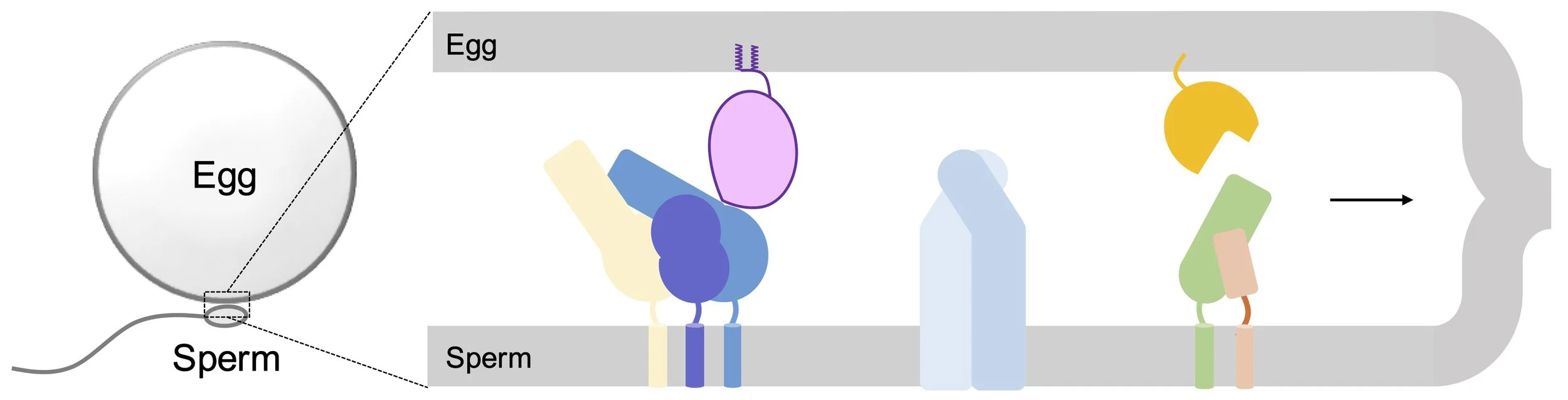
Our ongoing research efforts are broadly divided into two parts: first, we focus on identifying and characterizing new egg-membrane receptors in fertilization. We aim to understand how egg receptors define cell-surface recognition and species specificity. Second, we are interested in determining the molecular architecture of the sperm membrane proteins that are essential for fertilization. We aim to uncover the functional organization of the sperm surface proteomes and the molecular mechanisms underlying egg-sperm adhesion and membrane fusion. Understanding these molecular intricacies represents a basic science question with broad implications for human fertility and reproductive health.


Additionally, we are interested in cell–cell membrane fusion processes during human development, viral infection, and host-parasite interactions. We aim to investigate the diverse membrane-fusion machines at the levels of cell biology, mechanistic biochemistry, and structural biology. Our long-term goal is to exploit the structural and mechanistic information to design and develop new therapeutics and vaccines against infectious diseases.
Key publications
Liu, P., Castelino, R.E., Gierke, T.R., Wood, A.J., Lu, Y., Tang, S. (2026) Structure and inhibition of the sperm TMEM95-FIMP complex in mammalian fertilization. bioRxiv 2026.05.14.724122. PMCID: PMC13192804. PMID: 42182310. [PDF] [Github]
Lu, Y., Ikawa, M., Tang, S. (2025) Allosteric inhibition of the IZUMO1-JUNO fertilization complex by the naturally occurring antisperm antibody OBF13. Proc Natl Acad Sci U S A 122(10):e2425952122. PMCID: PMC11912406. PMID: 40042902. [PDF]
Lu, Y., Shimada, K., Tang, S., Zhang, J., Ogawa, Y., Noda, T., Shibuya, H., Ikawa, M. (2023) 1700029I15Rik orchestrates the biosynthesis of acrosomal membrane proteins required for sperm–egg fusion. Proc Natl Acad Sci U S A 120(8) e2207263120. PMCID: PMC9974436. PMID: 36787362. [PDF]
Tang, S., Lu, Y., Skinner, W.M., Sanyal, M., Lishko, P.V., Ikawa, M., Kim, P.S. (2022) Human sperm TMEM95 binds eggs and facilitates membrane fusion. Proc Natl Acad Sci U S A 119 (40) e2207805119. PMCID: PMC9546558. PMID: 36161911. [PDF]
Chang, H., Gierke, T.R., Tang, S., Lu, Y. (2026) Molecular interplay between sperm and egg: a narrative review. Hum Reprod Update. Online ahead of print. PMID: 41884958. [PDF]
Engineering cell-surface receptors and their ligands for therapeutics discovery and vaccine design
Recognition and signaling through cell-surface receptors and their ligands have designated them as promising clinic targets. We are interested in identifying new cell-surface receptor/ligand interactions and designing tailor-made therapeutics against the receptor/ligand targets in cancer and infectious diseases.

We are interested in targeting traditionally “undruggable” protein surfaces, where identifying small, drug-like molecules have failed empirically. For example, small-molecule immune checkpoint drugs have the potential to offer efficacy, safety, and affordability for cancer patients, but such inhibitors have been out of reach. We identified a prominent pocket on the immune checkpoint PD-1 protein, which has since become an attractive drug target for cancer immunotherapy. The pocket forms when PD-1 is bound to its native ligand, PD-L2. We observed allostery between the pocket and two adjacent loops of PD-1, opening new avenues for the discovery of small-molecule immunotherapy.
We are interested in developing methods to identify ligands for “undruggable” protein surfaces. For example, a conserved, hydrophobic pocket found in the HIV-1 envelope gp41 is a target for blocking viral entry. But it has proven challenging to target this pocket for eliciting neutralizing antibodies via immunization. We uncovered conformational flexibility in the gp41 pocket and designed peptide mimetics to rigidify its structure. We found that such peptides can elicit high avidity antibodies against the pocket and a modest neutralizing response against HIV-1. We propose that conformationally stabilized immunogens could serve as the basis for targeting “undruggable” sites during viral membrane fusion.

Additionally, we are interested in devising protein-based therapeutic and vaccine approaches against infectious agents. For example, we designed a ferritin nanoparticle vaccine displaying the SARS-CoV-2’s Spike protein. When formulated with alum adjuvant, this vaccine elicited high-titer neutralizing antibodies against major SARS-CoV-2’s variants of concern in animal models. This demonstrates how multivalent presentation of viral antigens can drive a robust polyclonal response. In parallel efforts, we use epitope-focusing methods in rational vaccine design and protein language models in guiding antibody evolution in infectious diseases.
Key publications
Tang, S. and Kim, P.S. (2019) A high-affinity human PD-1/PD-L2 complex informs avenues for small-molecule immune checkpoint drug discovery. Proc Natl Acad Sci U S A 116, 24500-24506. PMCID: PMC6900541. PMID: 31727844. [PDF]
Filsinger Interrante, M.V., Tang, S., Kim, S., Shanker, V.R., Hie, B.L., Bruun, T.U.J., Wu, W., Pak, J.E., Fernandez, D., Kim, P.S. (2025) Utilizing Machine Learning to Improve Neutralization Potency of an HIV-1 Antibody Targeting the gp41 N-Heptad Repeat. ACS Chem Biol. 20(7):1470-1480. PMCID: PMC12281478. PMID: 40540236. [PDF]
Bruun, T.U.J., Tang, S., Erwin, G.S., Deis, L.N., Fernandez, D., Kim, P.S. (2023) Structure-guided stabilization improves the ability of the HIV-1 gp41 hydrophobic pocket to elicit neutralizing antibodies. J Biol Chem 299(4):103062. PMCID: PMC10064241. PMID: 36841484. [PDF]
Weidenbacher, P.A., Sanyal, M., Friedland, N., Tang, S., Arunachalam, P.S., Hu, M., Kumru, O.S., Morris, M.K., Fontenot, J., Shirreff, L., Do, J., Cheng, Y., Vasudevan, G., Feinberg, M.B., Villinger, F.J., Hanson, C., Joshi, S.B., Volkin, D.B., Pulendran, B., Kim, P.S. (2023) A ferritin-based COVID-19 nanoparticle vaccine that elicits robust, durable, broad-spectrum neutralizing antisera in non-human primates. Nat. Commun. 14(1):2149. PMCID: PMC10110616. PMID: 37069151. [PDF]
Xu, D., Carter, J.J., Li, C., Utz, A., Weidenbacher, P.A., Tang, S., Sanyal, M., Pulendran, B., Barns, C.O., Kim, P.S. (2024) Vaccine design via antigen reorientation. Nat Chem Biol. 20(8):1012-1021. PMCID: PMC11247139. PMID: 38225471. [PDF]
Assembly and architecture of membrane-remodeling machines
Cell-surface receptors tune their signaling by internalizing themselves in the endo-lysosomal pathway. Receptors destined for recycling and lysosomal degradation are sequestered and packaged at endosomes by the endosomal sorting complexes required for transport (ESCRT). ESCRTs are conserved hetero-oligomeric machines that catalyze membrane fission reactions. ESCRT induces membrane budding away from cytoplasm, a topology shared among processes in multivesicular endosome biogenesis, HIV-1 budding, cytokinesis, etc.

We are interested in how the molecular machines, like the ESCRT-III complex, assemble and execute their membrane-sculpting functions. We determined the first core structures of the ESCRT-III complex, revealing that ESCRT-III activation requires a prominent conformational rearrangement that promotes the assembly of a membrane-bound complex. We identified that ESCRT-III interacts with lipids through hydrophobic membrane-insertion motifs, which facilitate the ESCRT-III’s ability to induce membrane invagination. The upstream ESCRT complexes, ESCRT-I/-II and ESCRT-0/Bro1, assist ESCRT-III’s assembly through ubiquitin-dependent receptor sorting. We showed that cooperative interactions between subunits within the ESCRT-III polymer enable architectural changes of ESCRT-III at distinct stages of membrane deformation. Collectively, our work defined the molecular mechanisms governing the activation, assembly, and membrane binding of the ESCRT-III complex in a spatially unique membrane fission reaction in the cell.
Key publications
Tang, S., Henne, W.M., Borbat, P.P., Buchkovich, N.J., Freed, J.H., Mao, Y., Fromme, J.C., and Emr, S.D. (2015) Structural basis for activation, assembly and membrane binding of ESCRT-III Snf7 filaments. eLife 4. PMCID: PMC4720517. PMID: 26670543. [PDF]
Tang, S., Buchkovich, N.J., Henne, W.M., Banjade, S., Kim, Y.J., and Emr, S.D. (2016) ESCRT-III activation by parallel action of ESCRT-I/II and ESCRT-0/Bro1 during MVB biogenesis. eLife 5. PMCID: PMC4865371. PMID: 27074665. [PDF]
Banjade, S., Tang, S., Shah, Y.H., and Emr, S.D. (2019) Electrostatic lateral interactions drive ESCRT-III heteropolymer assembly. eLife 8. PMCID: PMC6663469. PMID: 31246173. [PDF]
Banjade, S., Shah, Y.H., Tang, S., and Emr, S.D. (2021) Design principles of the ESCRT-III Vps24-Vps2 module. eLife 10. PMCID: PMC8143795. PMID: 34028356. [PDF]
Buchkovich, N.J., Henne, W.M., Tang, S., and Emr, S.D. (2013) Essential N-terminal insertion motif anchors the ESCRT-III filament during MVB vesicle formation. Dev Cell 27, 201-214. PMID: 24139821. [PDF]